
Therapeutic Areas
Kidney Disease
Drug Discovery Services
Axcelead supports chronic kidney disease (CKD) and acute kidney injury (AKI) drug discovery with characterized disease models, clinically relevant endpoints, and integrated molecular analysis. Our pharmacology team helps you select appropriate models, evaluate preclinical treatment effects, and interpret drug action in kidney tissue.

THE RESEARCH CHALLENGE
Selecting the right model and understanding the response
Kidney diseases vary in disease stage, affected nephron compartments, fibrosis, inflammation, and the balance of glomerular and tubular injury. A single model cannot capture this diversity. The kidney also contains multiple cell populations with distinct disease responses, making it important to understand where and how a treatment acts. Model selection, endpoint selection, and tissue analysis need to address the specific questions in your program.
OUR APPROACH
How Axcelead supports your program
Explore three capabilities that connect model selection, preclinical evaluation, and mechanistic interpretation.
Backed by extensive experience in CKD and AKI pharmacology, we connect model selection with functional, biomarker, molecular, and histopathological evaluations to address your research question.
A diverse portfolio of kidney disease models
Our portfolio includes CKD and AKI models. CKD models cover genetic and multifactorial disease, nephron loss, immune-mediated glomerular injury, tubulointerstitial injury, diabetic kidney disease, and renal fibrosis. AKI models cover ischemia-reperfusion and cisplatin-induced injury. We help you select a model that matches the disease process and study objectives.
View the model portfolio overview
CKD models
- AXCC mouse
- SHC rat
- 5/6 nephrectomized rat
- Glomerulonephritis Rat
- Adenine-loaded mouse
- Unilaterally nephrectomized db/db mouse
- Unilateral ureteral obstruction (UUO) mouse
AKI models
- Ischemia–reperfusion injury mouse
- Cisplatin-induced AKI mouse
Model selection and availability can be discussed with the Axcelead pharmacology team.
Compare characteristics of CKD models
| Models | Disease mechanism | Albuminuria | GFR decline | Fibrosis | Key feature |
|---|---|---|---|---|---|
| CKD patients (advanced stage) | Multifactorial | Marked | Consistent | Marked | – |
| AXCC mouse | COL4A5 mutation | Marked | Consistent | Marked | Alport syndrome |
| SHC rat | Multifactorial | Marked | Consistent | Marked | Focal segmental glomerulosclerosis (FSGS) |
| 5/6 Nx rat | Nephron loss | Moderate | Variable | Marked | Hyperfiltration |
| Glomerulonephritis model | Anti-GBM antibody | Marked | Variable | Moderate | Crescent formation |
| Adenine-loaded mouse | Tubular injury | Low | Variable | Moderate | Tubulointerstitial nephropathy |
| Unilateral Nx db/db mouse | Metabolic and nephron loss | Moderate | Variable | Moderate | Diabetic kidney disease (DKD) |
| UUO mouse | Urinary tract obstruction | N/A | N/A | Marked | Fibrosis |
Table 1. Qualitative characteristics of CKD models. Advanced-stage CKD patients are included as a clinical reference, separate from the seven animal models. Descriptors depend on the model and study conditions and do not represent a head-to-head comparison. N/A: not applicable in this comparison.
Measure kidney function and disease-associated biomarkers
Available assessments include non-invasive GFR measurement, urinary albumin and protein, and blood creatinine and urea nitrogen, enabling longitudinal monitoring of renal function and disease progression.
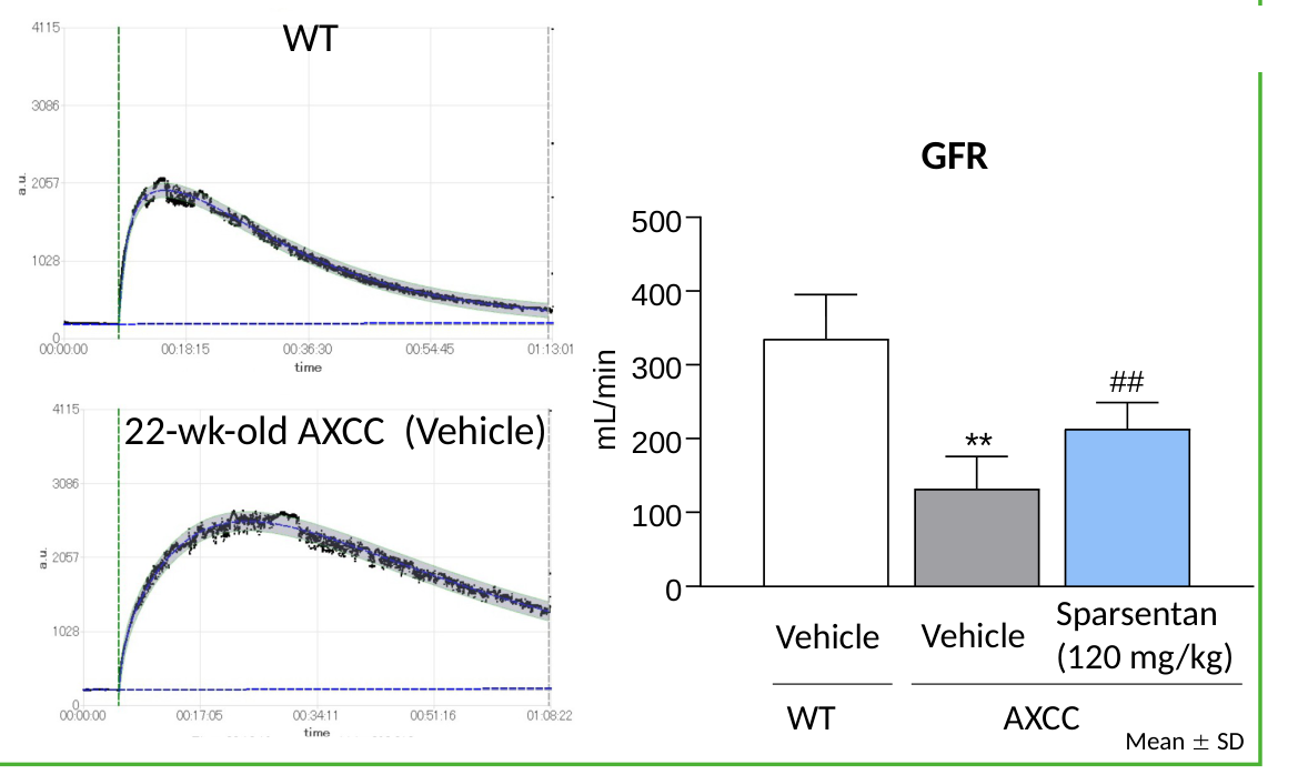
Among these assessments, non-invasive GFR measurement enables repeated evaluation of renal function in the same animal throughout the study. The example below demonstrates longitudinal GFR assessment in the AXCC CKD model and pharmacological improvement following chronic treatment.
In addition to standard measures of renal function and injury, such as urinary albumin, proteinuria, blood creatinine, and urea nitrogen, a broad range of biomarkers can be incorporated to evaluate specific disease processes. Representative examples are shown below.
| Disease process | Example biomarkers |
|---|---|
| Reduced kidney function | Cystatin C |
| Tubulointerstitial injury | NGAL, KIM-1, L-FABP |
| Glomerular injury | Podocin, nephrin |
| Endothelial dysfunction | ADMA |
| Oxidative stress | TBARS, 8-OHdG, 4-HNE |
| Inflammation | Cytokines, CRP |
| Fibrosis | TIMP-1 |
Readouts are tailored to the model and study objectives; the examples above do not represent a fixed assessment panel for all models.
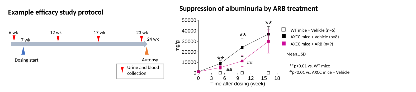
AXCC mice and SHC rats are proprietary CKD models with translational relevance to progressive kidney disease. Their well-characterized phenotypes and pharmacological responses support robust evaluation of disease progression and treatment effects, helping guide study design and assessment strategies.
AXCC mouse: Alport syndrome and progressive CKD
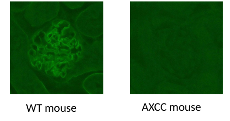
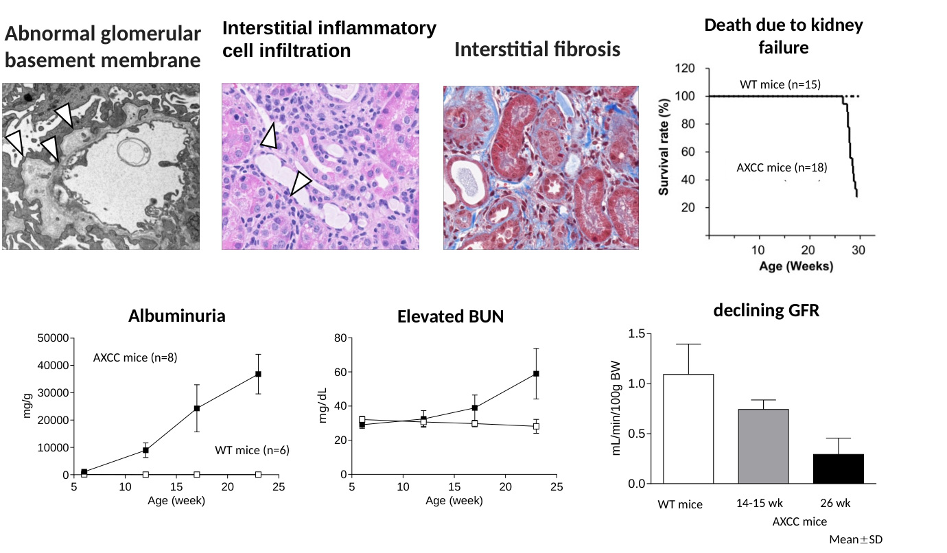
Developed by Axcelead, AXCC (Axcelead DDP-generated Col4a5 R471* chronic kidney disease model) carries the Col4a5 R471* mutation identified in people with Alport syndrome. This genetically defined model recapitulates key pathological features of Alport syndrome and progressive CKD, supporting preclinical efficacy assessment and translational research. Glomerular COL4A5 loss and basement membrane abnormalities are accompanied by progressive albuminuria, interstitial inflammation, fibrosis, and kidney dysfunction.
- Low inter-animal variability and reproducible disease progression and pharmacological responses support consistent preclinical evaluation.
- Characterized responses to clinically effective therapies, including angiotensin II receptor blockers (ARBs) and dual endothelin type A/angiotensin II receptor antagonists, support the translational relevance of the model.
- Existing spatial transcriptomics data cover more than 20,000 genes. Together with single-cell RNA-sequencing data, these datasets enable evaluation of target expression and disease-associated pathways before study initiation; analytical detail is provided in Approach 3.
- Supply of more than 80 AXCC mice per study supports large-scale efficacy evaluation and statistically informed study design. Availability and sample size are confirmed during study planning.
The AXCC model exhibits a reproducible sequence of disease progression, providing defined study windows for the evaluation of renal injury, functional decline, and therapeutic response.
| Approximate age | Disease milestone |
|---|---|
| 7 weeks | Albuminuria |
| 14 weeks | Fibrosis |
| 17 weeks | Elevated BUN |
| 20 weeks | Decreased GFR |
| 25 weeks | Kidney failure-associated mortality |
Publications and scientific presentations
Peer-reviewed publications
- Podocyte specific exon skipping after disease onset improves kidney pathology and function in a mouse model of Alport syndrome.
Hashikami K, et al. | Scientific Reports. 2025;15:41766. - A Novel Deep Learning Approach for Analyzing Glomerular Basement Membrane Lesions in a Mouse Model of X-Linked Alport Syndrome.
Kawanishi K, et al. | American Journal of Pathology. 2025;195(1):143-154. - Bidirectional, non-necrotizing glomerular crescents are the critical pathology in X-linked Alport syndrome mouse model harboring nonsense mutation of human COL4A5.
Song JY, et al. | Scientific Reports. 2020;10:18891. - Establishment of X-linked Alport syndrome model mice with a Col4a5 R471X mutation.
Hashikami K, et al. | Biochemistry and Biophysics Reports. 2019;17:81-86.
Conference presentations
- Inducible exon skipping after disease onset as a therapeutic strategy for CKD: Evaluation in a patient-derived mouse model of Alport syndrome.
Hashikami K, et al. | World Congress of Nephrology, 2026 - Irbesartan, but not roflumilast, ameliorates renal injury in a Col4a5 R471 mutant mouse model of Alport syndrome and CKD.
Yamamoto M, et al. | World Congress of Nephrology, 2026 - Impact of timing on glomerular-specific restoration of COL4A5 expression in Alport syndrome model mice.
Hashikami K, et al. | Japanese Society of Nephrology, 2024 - Glomerular-specific restoration of COL4A5 expression in Alport syndrome model mice by the Cre-loxP system.
Hashikami K, et al. | Japanese Society of Nephrology, 2023 - Tubulointerstitial chronic lesions and tertiary lymphoid tissue in a mouse model of Alport syndrome.
Kobayashi R, et al. | Japanese Society of Nephrology, 2022 - Do the pharmacokinetic profile of Axcelead DDP-generated Col4a5 R471X-chronic kidney disease model (AXCC) mice reflect human renal disease?
Kato N, et al. | Japanese Society for the Study of Xenobiotics, 2022 - Phenotype analysis and efficacy of irbesartan in Col4a5 R471X mice harboring a mutation similar to an Alport syndrome patient.
Hashikami K, et al. | Molecular Biology Society of Japan, 2020
SHC rat: metabolic and hemodynamic abnormalities with FSGS-like injury
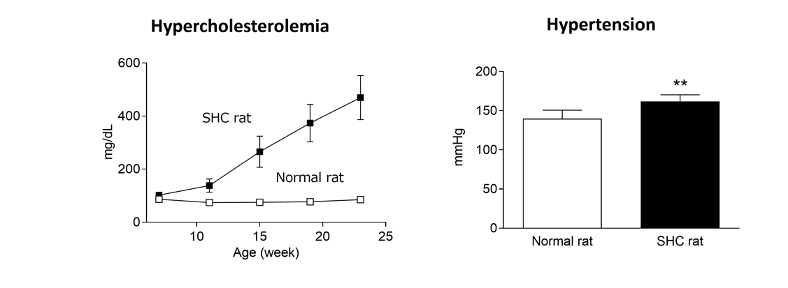
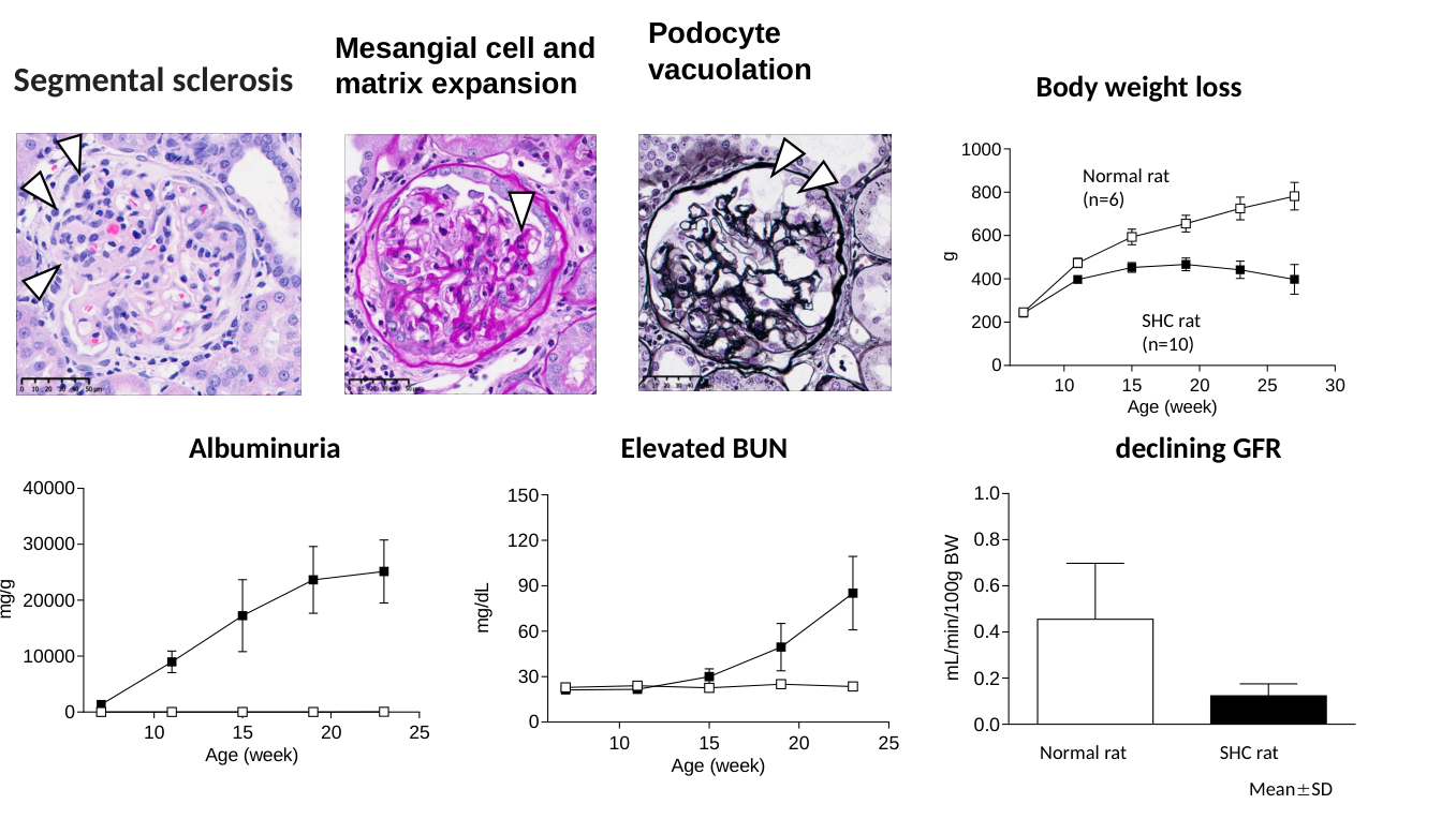
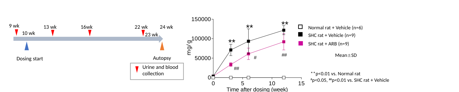
The spontaneously hypercholesterolemic (SHC or Imai) rat is a translationally relevant CKD model that reproduces key clinical features of progressive kidney disease. Spontaneous hypercholesterolemia and hypertension drive the development of glomerulosclerosis and FSGS-like lesions, resulting in progressive CKD with translational relevance to human disease. Disease progression is characterized by albuminuria, elevated BUN, reduced GFR, and kidney failure-associated mortality. Low inter-animal variability and highly reproducible disease progression and pharmacological responses support reliable efficacy assessment and mechanistic studies.
The SHC rat exhibits a reproducible sequence of CKD progression driven by metabolic and hemodynamic abnormalities, providing defined study windows for the evaluation of renal dysfunction and therapeutic response.
| Approximate age | Disease milestone |
|---|---|
| 7 weeks | Albuminuria |
| 15 weeks | Elevated BUN |
| 20 weeks | Decreased GFR |
| 25 weeks | Kidney failure-associated mortality |
Publications and scientific presentations
Conference presentations
- The SHC (Spontaneously Hypercholesterolemic) rat as a Translational Model of FSGS: Renoprotective Effects of Irbesartan.
Takagahara, et al. | Kidney Week, 2026
The kidney contains diverse, spatially organized cell populations. We integrate molecular profiling with pharmacology and histopathology to characterize drug effects and mechanisms of action in depth.
Technology platforms
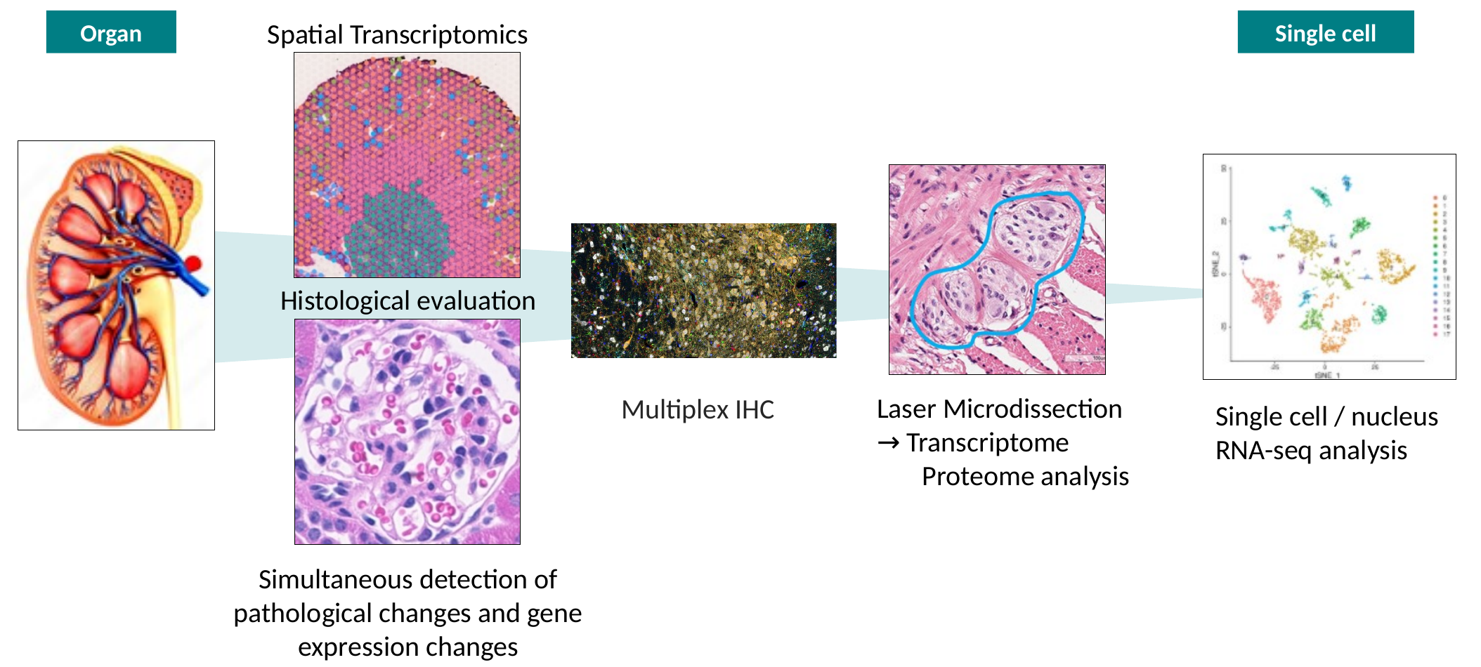
Technology platforms for integrated kidney tissue and molecular analysis.
Spatial transcriptomics
Simultaneous assessment of histopathology and genome-wide gene expression
- Localize drug-responsive molecular changes within kidney tissue
- Link molecular responses to histopathological lesions
Multiplex IHC
Multiparametric tissue imaging using simultaneous detection of multiple markers
- Characterize cellular composition and spatial organization of diseased tissue
- Quantify treatment-related changes in immune and resident cell populations
Laser microdissection
Selective collection of defined tissue regions for downstream molecular analysis
- Isolate specific renal structures or pathological lesions for targeted transcriptomic and proteomic analysis
- Link molecular changes to histopathological findings at the lesion level
Single-cell / single-nucleus RNA sequencing
Cell-resolved transcriptomic profiling at single-cell resolution
- Identify cell populations associated with disease and treatment response
- Characterize cell type-specific pathways and molecular responses
Case study 1: Spatial transcriptomics in AXCC kidneys
Key question: Where do disease-associated gene-expression programs emerge within the AXCC kidney?
Spatial maps across kidney sections
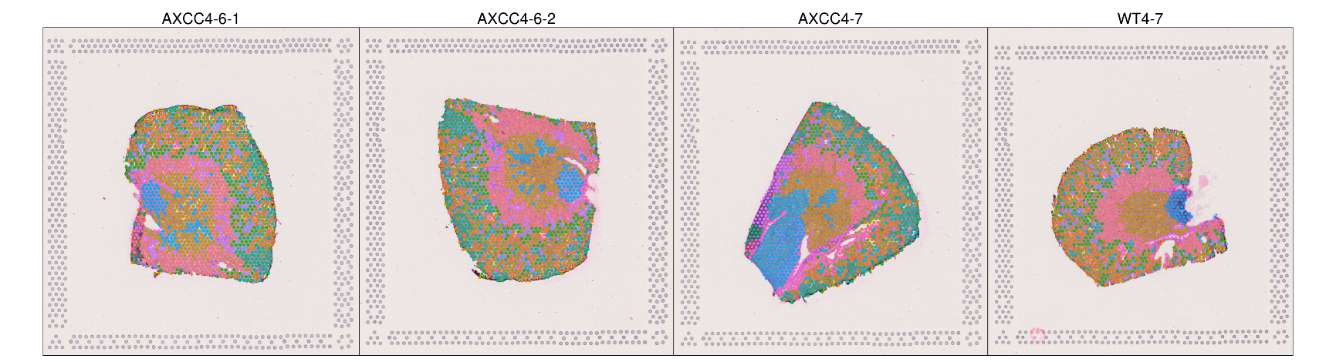
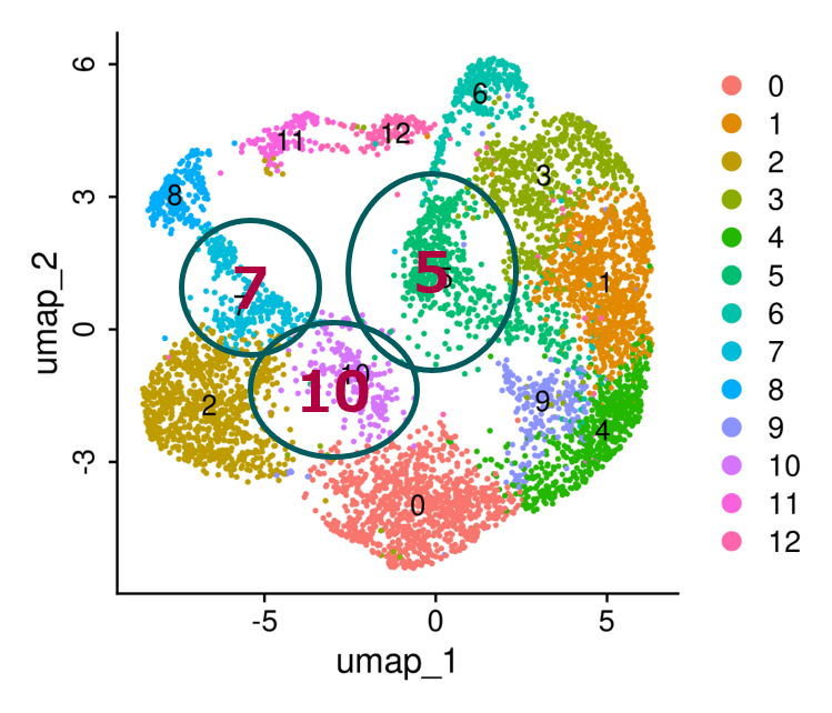
Gene-expression clustering
Interpretation
Clusters 5, 7, and 10 are enriched in AXCC kidneys, identifying spatially restricted disease-associated molecular programs.
Higher-resolution spatial analysis is also available at 2-µm resolution.
Key finding: Spatial profiling connects AXCC molecular signatures with the kidney regions in which disease pathology develops.
Case study 2: Single-cell RNA sequencing in AXCC kidneys
Key question: Which kidney cell populations and molecular pathways are most affected in AXCC disease?
Cell type-specific expression patterns
Differentially expressed genes distinguish kidney cell populations and support cell type annotation.
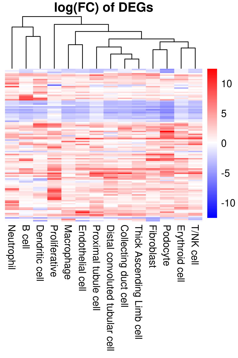
Cell population landscape
UMAP identifies cell populations associated with disease and treatment response.
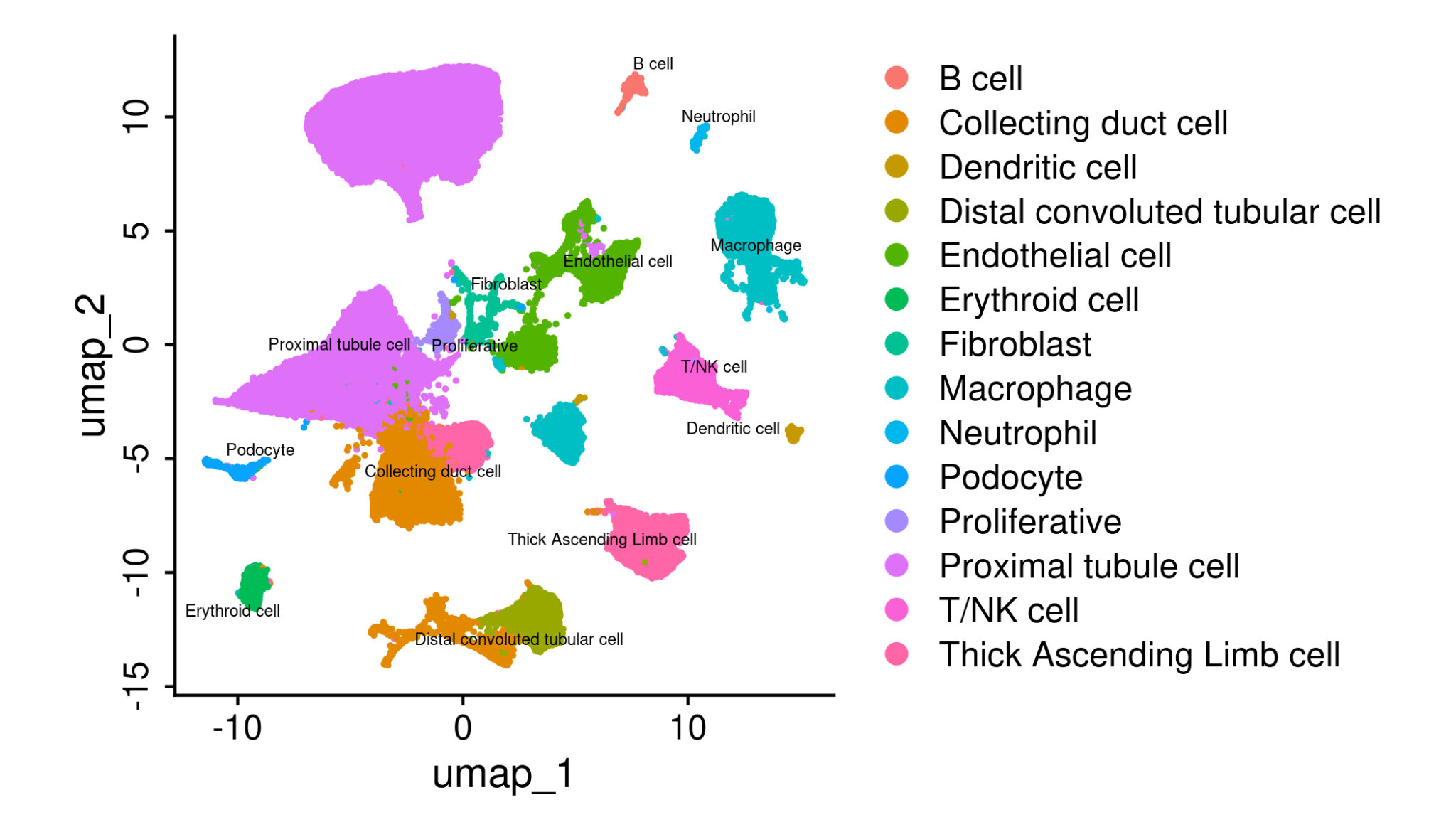
Key finding: Single-cell RNA sequencing characterizes cell type-specific transcriptional changes in AXCC kidneys and provides a resource for target and biomarker evaluation.
Supplementary information
Leverage existing AXCC molecular datasets before initiating in vivo studies
- Assess target expression before study initiation
- Explore tissue- and cell type-specific expression patterns
- Support selection of molecular endpoints and biomarkers